Doug Butler, Ph.D., CJF, FWCF defines hoof balance as a condition that exists when the weight placed on the legs of a horse is equally distributed over the foot of each leg. Veterinarians, farriers and equestrians have long recognized that a well-balanced equine hoof is a prerequisite to soundness and optimal performance for the equine athlete. The development of numerous pathologic conditions, both chronic and acute, have been attributed to poor hoof balance. Until recently, very little objective data have been available pertaining to correct hoof balance and the determination thereof. Hoof balance continues to be determined and achieved by utilizing anecdotal evidence, despite significant improvements in both veterinary and farrier sciences. The goal of this paper is to describe and explain hoof balance and to present scientific evidence from the past three decades. It is my hope that this information will assist you when evaluating equine patients.
Static Versus Dynamic Balance
One can evaluate hoof balance in two ways. Static balance considers the proportionality and geometry of the hoof at rest, bearing full body weight while the horse is standing squarely. Ideally, the animal is positioned on a smooth, firm, level surface during the evaluation. Dynamic balance requires observation of the horse while moving, so that the flight phase and stance phase of the limbs in motion can be ascertained. Again, use of a smooth, firm, level surface is considered ideal.
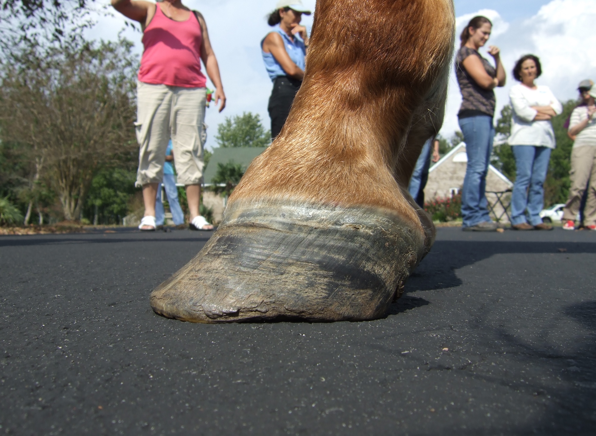
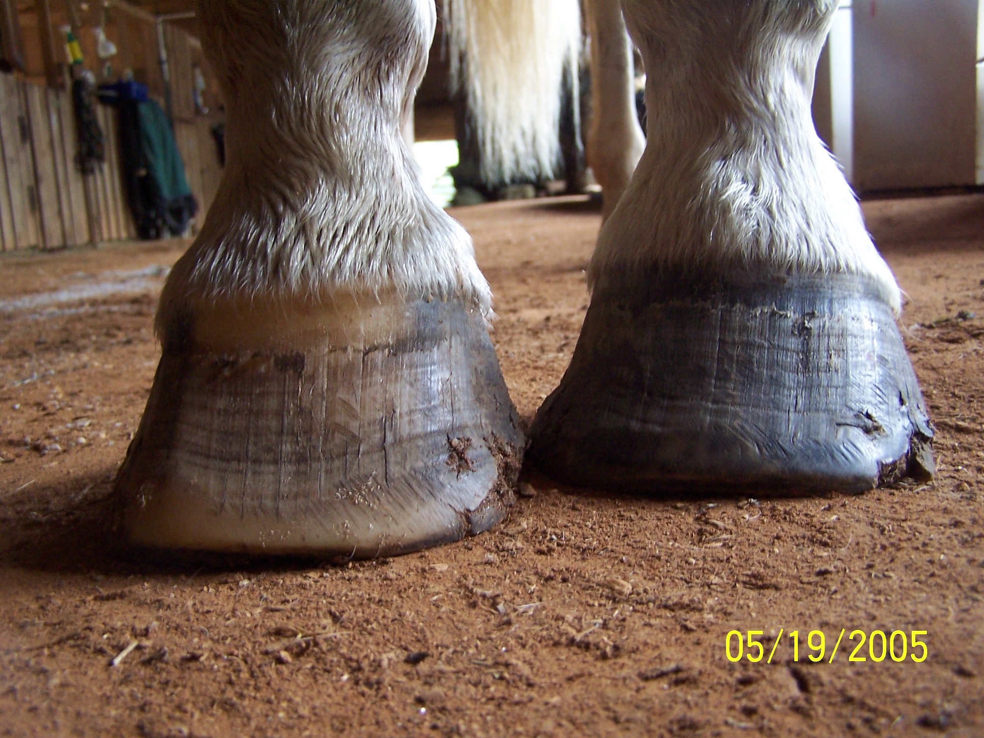
Static balance is most often assessed visually. Determination of hoof balance requires that we appreciate proportions and angles displayed by the hoof of interest. In general, a well-balanced hoof exhibits a straight hoof-pastern axis (HPA) such that when viewed from the side, the angles of the dorsal hoof wall and the pastern are the same. It has been suggested that the angles of the dorsal hoof wall and heels should be equal, and that heel length should be 1/3 that of toe length. The walls at the toe and the heel should be straight. Mediolateral balance exists when the lengths of the medial and lateral hoof walls results in the coronary band being parallel to the bearing surface (bottom) when viewed from the front, and the bearing surface is perpendicular to a line drawn through the center of the pastern and cannon bone. The walls should be straight, though the angles need not be identical. The hoof wall with the more acute angle will have a greater length, but this should not influence the orientation of the coronary band with respect to the bearing surface.

Methods to achieve static balance include using hoof gauges to measure angles, and various methods of mapping the hoof. (Left, THH files. Right, courtesy Gwenyth Santagate.)
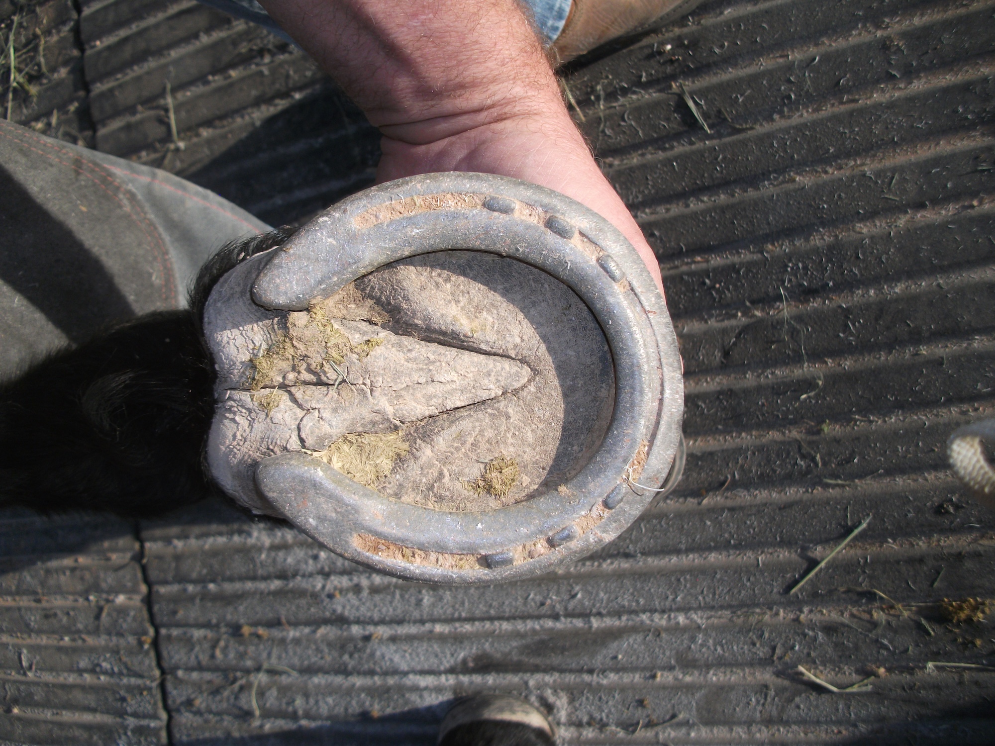
The bearing surface of the hoof capsule should be divided equally into medial and lateral halves. The width of the base of the frog should be 2/3 of the length of the frog, and the length of the frog should equal to 2/3 of the length of the bearing surface, as measured from the termination of the heels to the toe. The size of the hoof capsule should be in proportion to the size and mass of the horse, and contralateral hooves should be symmetrical. Objective measurements can be made using devices to determine hoof angle and length. Farriers and trimmers commonly use hoof gauges to measure the angle of the hoof wall before and after a trim.
Determining Hoof Balance
There are numerous methods used to determine static balance, and not all methods consider the same parameters. Geometric balance is determined visually as described above. This can be performed in the live horse, or by using photographs of the hooves. Critics of this method point out that very few horses can meet the criteria for “normal” balance, and consider the geometric approach invalid.
Dave Duckett developed a method to determine hoof balance based on the center of the hoof capsule. “Duckett’s Dot” serves as the reference point for this method, and it is used to locate the center of the bearing surface of the hoof. The point is located at the intersection of a line drawn across the widest part of the bearing surface and a line bisecting the center of the frog. This point is typically found between 1/2 and 1 inch behind the apex of the frog.
Diagonal hoof balance is similar to Duckett’s theory. In this method, lines are drawn from the heel to the opposite toe, and the point where the lines cross should be the center of the hoof. In both methods, the point represents the center of the hoof.
The natural balance and four-point trim methods are similar in that both suggest optimal balance is achieved when the hooves are trimmed to resemble those of feral horses. Proponents of both methods theorize that natural selection has determined ideal hoof balance through adaptation and survival.
The “hairline” method utilizes the shape of the coronary band as a reference point. When the hairline deviates from a straight line it is indicative of areas of excess stress on the bearing surface, resulting in raised or “jammed up” areas in the coronary band. Proponents suggest trimming the hoof wall to return the coronary band to proper orientation in order to achieve normal hoof balance.

Photo courtesy Epona
Radiographic evaluation is considered the gold standard for assessing hoof balance. A combination of lateromedial and dorsopalmar/plantar views are required, and computer software is available to measure and calculate the parameters with which we determine hoof balance.
All of the techniques described above enable us to make immediate “before and after trimming” comparisons of the hooves. Our goal is to improve hoof balance with each intervention, and to have a baseline for future comparisons.
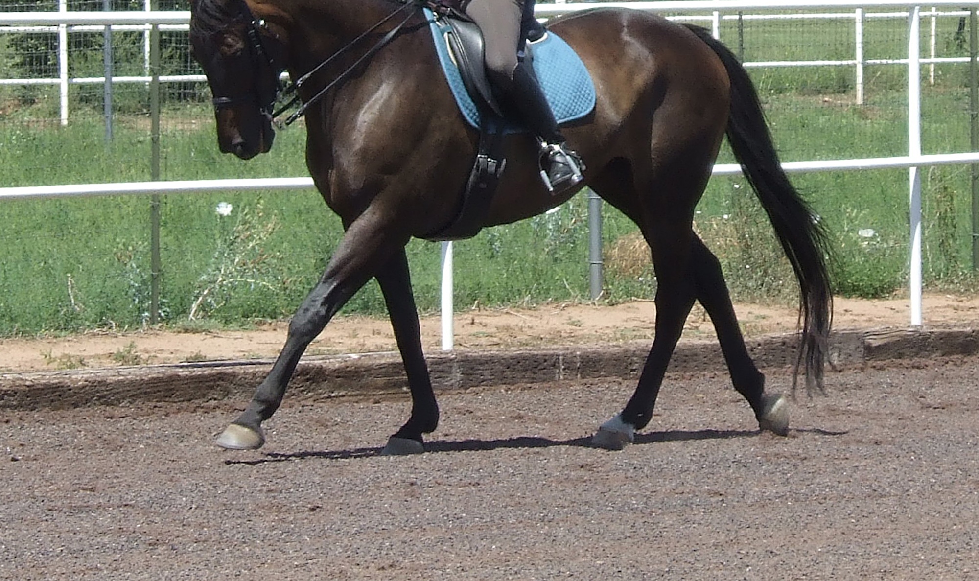
In order to achieve dynamic balance, the horse must be observed while moving. Trimming and shoeing intervention is performed in an attempt to alter or otherwise improve the balance of the limbs with regard to foot flight, landing, and take off. The goal is to balance loading and unloading of the hooves to minimize abnormal stresses and improve mechanical efficiency. Most hoof care providers and veterinarians believe that the hoof should land level (medial and lateral sides contact the ground simultaneously) and either flat or slightly heel-first. Weight should be borne evenly across the bottom of the hoof during the stance phase, and break over or toe-off should occur at the center of the toe. Any deviation is considered less than ideal and potentially injurious to the hooves and limbs. Dynamic balance assessment can be performed by direct visualization of the horse in motion, or by using recording devices. The horse is observed from the front, sides, and rear while moving. More recently, pressure-plate analysis of the feet has been used to objectively measure dynamic balance.
Can We Accurately Determine and Achieve Hoof Balance?
Moleman et al. conducted a study to determine the accuracy of different hoof gauges (1). Six individuals using four different hoof gauges each measured dorsal hoof wall angle in 10 sound horses. Results were compared to measurements derived from digital radiographs of each hoof. The devices were tested for reliability and validity after training each operator in the proper use of each device. The researchers found that the size and shape of the hooves led to different measurements, and that hoof angle measured using the same device varied among hooves and operators such that comparisons could not be made. The operators used the gauges differently despite identical training. The authors concluded that no device was both reliable and valid, and none was very accurate.
Radiographic determination of hoof balance, long considered the gold standard, is also subject to error and inaccuracy. Researchers evaluated the effect of patient position on accuracy of measurements to determine balance and angles in distal limb radiographs (2). They found a strong linear relationship between the horse’s stance and the hoof-pastern axis on lateromedial radiographs, up to a difference of 10 degrees, based on limb position. The dorsal angle of the distal phalanx was not influenced by stance. On dorsopalmar radiographs, a wide range of motion of the distal interphalangeal (DIP), but not the proximal interphalangeal (PIP), joint was found when stance changed; a wider stance altered the angle of the DIP joint resulting in an artificial imbalance. The study underscores the need for consistent positioning when comparing radiographs of the distal limbs. Proper positioning and consistent elevation of all four hooves is mandatory for meaningful radiographs.
A recent study attempted to validate a widely accepted trimming protocol that purportedly results in geometric balance when performed routinely (3). The investigators used a geometric balance based protocol to determine whether hoof balance is achieved in cadaver limbs (n=49) and in shod (n=6) or unshod (n=20) horses through three trimming cycles at five week intervals. The group found that this commonly used theory of geometric proportions for hoof balance did not hold true. Results lead them to question commonly accepted measures of ideal hoof balance such as dorsal hoof/heel parallelism, and the 3:1 ratio of dorsal wall to heel length (74 to 91% of study horses fell outside these ideals, even after three trimming cycles). Furthermore, their results question the current concepts of a standardized model of hoof balance that is applicable to all horses. The group’s conclusion was that hoof trimming should be done on an individual basis rather than trimming all feet to one ideal hoof model.
How Do Horses Actually Move?
The following investigations used pressure-plate analysis to determine symmetry in force distribution in limbs as well as pressure distribution over the contact area of the hoof. However, pressure plates cannot be used to measure absolute values of limb loading because of limitations in accuracy.
Van Heel and colleagues investigated dynamic loading of both front feet and hind feet in 18 clinically sound warmblood horses (4). The group found that lateral asymmetrical landing (outside hoof wall contacts the ground before inside hoof wall, at landing) was the preferred way of landing in the front feet, and especially the hind feet, of trotting horses. Corrective trimming, aimed at complete symmetry (outside and inside hoof walls of equal length) under static conditions, did not change this preference. They also found that lateral landing of trotting horses is not visible to the human eye. The group concluded that lateral asymmetrical landing is normal for warmblood horses, and that lateral rather than symmetrical landing should be regarded as the physiological standard (“normal”) for these horses.


With a force plate, the horse strikes a flat piece of metal that measures force of weight-bearing. Shown here, horses standing and trotting on two force plates with reflective markers attached to measure forelimb joint movements with kinematics and electromyography to measure forelimb muscle activation. Photos courtesy of Dr. Kevin Haussler, Colorado State University Equine Orthopedic Research Center.
A similar study investigated toe-heel and mediolateral hoof balance of sound sport horses at the walk and trot (5). Their hypothesis was that sound sport horses present a high degree of symmetry in hoof balance between contralateral fore limbs. Seven horses were led at a walk and trot over a pressure-force plate combination. The study determined that toe-heel balance presented a high degree of symmetry between contralateral fore limbs. Initially, the heel was loaded, quickly followed by toe loading. Loading became equivalent until 60-65% of stance, when toe loading increased. This pattern was the same for both limbs.
With respect to mediolateral balance, gait had an unexpected influence on the results. At the walk, both left and right fore feet exhibited higher loading of the lateral aspect of the hoof throughout the stance phase. At the trot, the left limb mediolateral balance presented higher loading of the medial part of the hoof at impact, whereas the right limb showed higher loading of the lateral part of the limb in all horses. At mid-stance and end of stance phases, there was no difference between rights and left; both limbs exhibited higher loading of the lateral part of the hoof at the end of stance. The authors were surprised by the difference that existed between contralateral limbs at the trot, but not the walk, and discussed motor laterality (left vs. right handed) and handler influence as possible explanations for their findings. However, it was determined that only two of the seven horses had left-handed preferences. The other five exhibited no preference. The authors repeated the study later with the handler positioned on the opposite side of the horse, but the results did not change. The authors made the following comment in the discussion section: “The paradigm of locomotor symmetry should be approached with caution.”
Oosterlinck and colleagues objectively evaluated toe-heel and mediolateral hoof balance of the vertical ground reaction force and limb-loading symmetry in five clinically sound, toed-in warmbloods (6). Toe-heel balance revealed higher loading of the toe zone at impact in 4/5 horses (toe-first landing?), while the remaining horse exhibited higher loading of the heel zone at impact. At mid-stance, loading of the toe and heel zones was equal in all horses. Towards the end of the stance phase, all five presented progressive loading of the toe zone. Mediolateral balance was similar in all horses with higher loading of the lateral zone at impact, equivalent loading at mid-stance, and increased loading of the lateral zone at the end of the stance phase. Investigators were uncertain as to why 4/5 horses loaded the toe region more than normal horses during the early stance phase, but speculated that it could represent subclinical disease or either cause OR effect of subclinical lameness. The authors acknowledged that the study was limited by the small number of horses, and the fact that horses were only evaluated at the walk.
The same group performed a study to determine the effect of a hard versus soft surface on dynamic hoof balance (7). They hypothesized that hoof loading and balance at the walk and trot would be different on a hard surface vs. a soft surface. Five ponies were evaluated over a hard surface (rubber mat) and a soft surface (sand and arena footing, 5 cm deep). The study documented a decrease in stance-phase duration and limb loading, and more equal force distribution between the toe and heel region and the medial and lateral hoof zones on a deformable surface, especially at impact. The investigators believed these differences were due to deformation of the sand surface on impact, resulting in additional energy dissipation. The ponies also exhibited a higher stride frequency in sand due to a shortened stride length, which resulted in a shorter stance phase and decreased loading. The authors concluded that surface characteristics affect dynamic hoof balance. (Who would have guessed?).
In 2014, Wilson et al published a study investigating the relationship between foot conformation, foot placement, and movement asymmetry (lameness) (8) . Foot conformation from photographs, foot placement at the walk and trot (photos) and movement asymmetry (lameness) of the front limbs was examined in 43 horses (33 ridden, 10 in hand) at the walk and trot. All horses were in work and considered sound by the riders/owners. The investigators reported the following findings:
- Twenty-two of 43 horses were sound (no evidence of movement asymmetry), 25.6% (11) were lame in the left front and 23.3% were lame in the right front.
- The majority of foot conformation parameters measured showed a significant degree of asymmetry within a hoof, suggesting that a geometrically balanced foot is rare in this group.
- Significant differences between the left front and right front feet were found in 50% of the hoof conformation parameters measured.
- Increasing amounts of movement asymmetry were related to shorter and/or narrower hooves (small feet), BUT they could not determine whether small feet were the cause or effect of lameness.
- The most common foot placement was lateral heel at the walk, and lateral at the trot.
- Fewer than half the horses showed consistent foot placement (defined as same foot placement 75% of the time).
- There were no differences in predominant placement of either the left or right at the walk or trot.
- Only the dorsal and palmar hoof angles had a significant impact on foot placement. A more acute (lower) angle was significantly associated with a lateral toe placement at the trot. None of the other conformation parameters was associated with foot placement at the walk or trot.
- There was no association between predominant foot placement and movement asymmetry at the walk or trot.
The investigators reached the following conclusions:
- Shorter, narrower hooves are related to movement asymmetry.
- Foot placement is independent of both movement asymmetry and, to a large extent, conformation.
- Horses may have a preferred or inherent way of placing their feet that supersedes the influence of conformation and movement asymmetry.

What is the Impact of Hoof Care on Hoof Balance?
To determine the effects of trimming on conformational parameter measurements, Kummer et al. conducted a study using 40 warmblood horses (9). Nine weeks after the most recent trim and shoe reset (all horses were shod) two radiographs were taken of both front feet. The feet were trimmed and radiographed again and shoes were applied. Eight weeks later, the process was repeated. The authors reported the following findings:
- A distinct degree of anatomical asymmetry was noted, as the dorsal hoof wall length was longer in right front hooves 65 to 77.5% of the time in the four different sessions.
- Radiographically, the digital bones and hoof capsule were significantly larger in the left front than the right front (70% of the time).
- Trimming significantly changed the hoof capsule conformation, especially in the toe region. Normal wear in the heels and minimal wear in the toe region allows a change in hoof wall length and hoof/P3 angle over time. Trimming significantly changed this.
The authors reached the following conclusions:
- The study showed that the trimming procedure has a large influence on hoof conformation, and that the changes in conformation observed after trimming could explain irregularities of gait shortly after trimming or shoeing (short, choppy gait).
- They theorized that trimming causes an abrupt alteration of loading patterns in joints, tendons and ligaments and that a period of adaptation to the new conformational parameters is required before normal gait is restored.
- They determined that mediolateral orientation of the boney structures of the hoof could not be determined by examining the hoof capsule. Only radiographic evaluation allowed accurate assessment of mediolateral orientation.
Moleman et al conducted a study to measure the effect of change in hoof conformation during a shoeing interval on the moments about the proximal and distal interphalangeal joints and to determine whether and how the horse compensates for this change in hoof conformation (10) . The study was performed using both front feet of nine warmblood horses during an eight-week shoeing interval. Findings included the following:
- Asymmetry between right front and left front was common, with one foot considered “low” hoof angle and the other one “high” hoof angle.
- Over time, feet became “broken back” and phalangeal alignment changed.
- This resulted in a change in the angle of the DIP joint (hyperextension), but not the PIP joint.
- The change in the DIP joint angle accommodates the change in hoof conformation, but results in greater tension on the deep flexor tendon, which affects the navicular area.
The authors reached the following conclusions:
- Dorsal hoof angle decreases during a shoeing interval of eight weeks.
- Horses compensate for this change by breaking the hoof-pastern axis backwards, thereby increasing DIP joint extension.
- This change is significantly greater in the “low” hoof angle foot.
- THUS, shoeing intervals should be kept short and should be individualized to the horse; the hoof with the lowest hoof angle is the best indicator as to when the horse should be reshod.
A similar study looked at the changes associated with the center of pressure and hoof un-rollment pattern over the course of an eightweek shoeing interval (11) . Eighteen sound warmblood horses shod by two different farriers (9 horses each) on an eight-week interval were used. The center of pressure (COP) was determined for each of the horse’s four feet, and hoof un-rollment patterns were recorded for each foot. The investigators reported these findings:
- After trimming and shoeing, the COP was most often located in the mediodorsal quadrant in the front feet, while in the hind feet the highest frequency was seen in the laterodorsal quadrant.
- After eight weeks, the COP moved in a palmar/plantar direction. In the front feet, mediolateral position of COP did not change significantly. In the hind feet, the preference for the laterodorsal quadrant became stronger.
- The hoof un-rollment pattern in the front feet changed little with respect to direction (dorsal), but the hind feet tended to shift the un-rollment pattern towards the lateral side (adaptive change?).
The authors reached the following conclusions:
- The horse can adapt to changes in hoof conformation over time, especially in the hind feet. A shift towards more lateral un-rollment appears to play an important role.
- Compensation may be most difficult when the hoof is altered by trimming at eight weeks due to an abrupt change in foot conformation.
What Impact Does the Hoof Care Provider Have on Hoof Balance?
Kummer et al performed a study to compare the effects of different farriers on radiograph-derived measurements of hoof balance (12) . This study was a spin-off of a previous study, (ref. #9). Forty horses were trimmed and shod by six qualified farriers, each of whom shod the same six or seven horses for the duration of the study. The shoes were removed, the feet radiographed, trimmed, and then radiographed again. Shoes were then reapplied. The entire process was repeated eight weeks later. Findings included the following:
- Despite the rather homogeneous group of horses, there were significant differences between farriers after two consecutive trimming procedures. In some cases, the differences were remarkable.
- Fourteen of 15 parameters that were influenced by the trimming procedure showed significant differences between farriers.
- Comparing the results of both trimming procedures revealed significant differences for several farriers, resulting from inconsistent technique between the two trimming procedures (different results from the same farrier trimming the same horse a second time).
- One farrier showed NO significant difference between sessions.
- In contrast, another farrier showed significant differences for four measured parameters between sessions.
The authors concluded:
- One goal of the trimming procedure should be to achieve a constant geometric balance and conformation of the digit.
- There were significant differences for most of the measured hoof parameters between the six farriers and between consecutive trimmings by the same farrier.
What Does the Horse Want?
Gordon and colleagues investigated the lower limb and hoof conformation of a population of semi-feral Mongolian horses (13) . The horses were used for riding, but had no routine hoof care of any kind, and were maintained on an open tundra environment. The study looked at 104 horses entered in the Mongolian Derby, a 1,000 km horse race. With respect to the hooves, the authors reported the following findings:
- Seventy-five percent of the horses displayed symmetry of size and balance of the front feet, while 25% were described as having uneven front feet.
- Hoof-pastern axis was consistently broken back, with a pastern angle of 60 degrees.
- As no owner reported providing routine hoof care or trimming, the hoof conformation in this group of Mongolian horses represented the natural interaction of the hoof with the environment.
The wild horse hoof has recently become a popular model for correct or natural hoof shape and balance, and numerous hoof care techniques attempt to emulate this model in domestic horse hooves. While reports of miraculous improvement in hoof conformation and resolution of lameness are plentiful, all information available thus far is purely anecdotal, and no hard scientific evidence has yet been presented to either support or refute these reports. Hampson has documented significant pathologic changes in the hooves of Australian brumbies, and has challenged the validity of the wild horse hoof as an ideal model for hoof care. Not all wild horse hooves have ideal conformation or balance, but these parameters appear to be more consistent among horses living in the same environment and sharing the same lifestyle. Indeed, the influence of diet, activity level, and environment may be more important than trimming technique, as the hoof has shown the ability to adapt and change in order to function optimally (14) .
My Thoughts
My advice echoes that of author Richard Carlson: Don’t sweat the small stuff. Understand that we created the concepts of perfect hoof balance and ideal hoof conformation. For more than a thousand years, we have attempted to dictate and mandate the shape and conformation of horse hooves without ever asking the horse for his opinion. Research conducted during the last three decades has dramatically improved our understanding of hoof function and biomechanics, but we are still far from being able to use that information to the betterment of the horse. My recommendations for equine hoof balance are based on years of experience with both lame and sound horses. Keep the toes short, keep the heels low, and keep the hoof walls straight.
Understand that hoof shape and conformation is often dictated by conditions you cannot change, such as conformation of the limb. Learn to recognize the impact of diet, activity and environment on hoof health, and seek to optimize these factors. Take the advice of Jaime Jackson, arguably the father of the natural hoof care movement in this country: First, do no harm; trust the healing powers of nature. I would add that if it ain’t broke, don’t fix it. Be patient. A single trim cannot change years of pathology. We are incapable of ‘fixing” a pathologic hoof, despite the significant strides that have been made in veterinary medicine and farriery. The best we can offer is to support the hoof and provide symptomatic therapy until the horse replaces the hoof with a newer, hopefully better hoof. This process can take years.
Use common sense. Never make sudden radical changes to the hoof. Trim frequently to prevent significant changes from developing in the first place. Hoof balance is a moving target; focus on the journey, not the destination. Good luck!
REFERENCES
- Moleman, M., Van Heel, M. C .V., Van Den Belt, A. J. M. and Black, W. (2005) Accuracy of hoof angle measurement devices in comparison with digitally analyzed radiographs. Equine Veterinary Education 17 (6), 319-322.
- Pauwels, F. E., Rogers, C. W., Wharton, H., Flemming, H., Wightman, P. F. and Green, R. W. (2017) Radiographic measurements of hoof balance are significantly influenced by a horse’s stance. Veterinary Radiology and Ultrasound 58 (1), 10-17.
- Caldwell, M. N., Allan, L. A., Pinchbeck, G. L., Clegg, P. D., Kissick, K. E. and Milner, P. I. (2016) A test of the universal applicability of a commonly used principle of hoof balance. The Veterinary Journal 207, 169-176.
- Van Heel, M. C. V., Barneveld, A., Van Weeren, P. R and Back, W. (2004) Dynamic pressure measurements for the detailed study of hoof balance: the effect of trimming. Equine Veterinary Journal 36 (8), 778-782
- Oosterlinck, M., Hardeman, L. C., van der Meij, B. R., Veraa, S., van der Kolk, J. H. and Wijnberg, I. D. (2013) Pressure plate analysis of toe-heel and medio-lateral hoof balance at the walk and trot in sound sport horses. The Veterinary Journal 198, e9-e13.
- Oosterlinck, M., Van der Aa, R., Van de Water, E. and Pille, F. (2015) Preliminary evaluation of toe-heel and mediolateral hoof balance at the walk in sound horses with toed-in hoof conformation. Journal of Equine Veterinary Science 35, 606-610.
- Oosterlinck, M., Royaux, E., Back, W. and Pille, F. (2014) A preliminary study on pressure-plate evaluation of forelimb toe-heel and mediolateral hoof balance on a hard vs. a soft surface in sound ponies at the walk and trot. Equine Veterinary Journal 46, 751-755.
- Wilson, A., Agass, R., Vaux, S., Sherlock, E., Day, P., Pfau, T. and Weller, R. (2016) Foot placement of the equine forelimb: Relationship between foot conformation, foot placement and movement asymmetry. Equine Veterinary Journal 48, 90-96.
- Kummer, M., Geyer, H. Imboden, I., Auer, J. and Lischer, C. (2006) The effect of hoof trimming on radiographic measurements of the front feet of normal warmblood horses. The Veterinary Journal 172, 58-66.
- Moleman, M., Van Heel, M. C. V., Van Weeren, P. R. and Back, W. (2006) Hoof growth between two shoeing sessions leads to a substantial increase of the moment about the distal, but not the proximal, interphalangeal joint. Equine Veterinary Journal 38 (2), 170-174.
- Van Heel, M. C. V., Moleman, M., Barneveld, A., Van Weeren, P. R. and Back, W. (2005) Changes in location of the centre of pressure and hoofunrollment pattern in relation to an 8-week shoeing interval in the horse. Equine Veterinary Journal 37 (6), 536-540.
- Kummer, M., Gygax, D., Lischer, C. and Auer, J. (2009) Comparison of the trimming procedure of six different farriers by quantitative evaluation of hoof radiographs. The Veterinary Journal 179, 401-406.
- Gordon, S., Rogers, C., Weston, J., Bolwell, C. and Doloonjin, O. (2013) The forelimb and hoof conformation in a population of Mongolian horses. Journal of Equine Veterinary Science 33, 90-94.
- Ramey, P. (2011). In: Care and Rehabilitation of the Equine Foot, Hoof Rehabilitation Publishing, Lakemont, GA USA.
About the author: Neal Valk, DVM, DACVS, CHCP is an equine veterinarian and board certified large animal surgeon in Greeneville, Tennessee. A graduate of the University of Tennessee, he has been in private practice for most of his career. He established the Stonehill Veterinary Center in Greeneville in 1998, and his special interest in natural hoof care began in 2004. He is currently Clinical Assistant Professor at University of Tennessee College of Veterinary Medicine. [in 2018]
Published in The Horse’s Hoof Magazine, Issue 70, Spring 2018
See the full content listing of all issues of The Horse’s Hoof Magazine! We also provide instructions on how to read the issues for free on Hoof Help Online.
For a detailed listing of all articles on The Horse’s Hoof website, please visit our Article Directory.