James & I attended Dr. Robert Bowker’s 2-day clinic in January, 2007, which was part of the Easycare-sponsored Pete Ramey & Dr. Bowker event. Though I had read about Bowker’s work for years, I had not grasped the scope or complexity of it, nor did I realize how compelling his collected evidence in support of barefoot is. THH Issue 26 included my quick summary of what I felt were some very intriguing—and controversial—points presented by Dr. Bowker. I hoped to whet people’s appetite, and spur them to find out more about what this amazing researcher has been doing.

Dr. Robert Bowker, January 2007.
Now I will attempt to delve deeper into my interpretation of what I saw presented by Dr. Bowker. Please read these ideas with an open mind, and understand that this is my perception. While you can disagree with interpretations and conclusions, they do not detract from the content of the actual scientific studies, which should really be examined first-hand.
Who is Dr. Robert Bowker?
Robert Bowker VMD PhD is a Professor of Anatomy and Director of the Equine Foot Laboratory at Michigan State Univ. College of Veterinary Medicine. His most important role (to us!) is that of veterinary researcher, and through funding support from the AQHA and Grayson Jockey Club, Bowker has been able to spend the last decade and a half studying the function of the equine foot. Much of his research has been on the microscopic level, and through the dissection and study of literally thousands of cadaver hooves.
The All-Important Back of the Foot
The rear of the hoof is possibly THE most important area for determining the health of the hoof. Bowker remarked that he kept trying to look elsewhere, but no matter what he did, it all kept coming back to the rear of the hoof!
The back of the foot is exactly where three of his most well-known studies have focused. The first was his hemodynamic flow theory, which proposes that blood flow through the network of tiny capillaries in the heel region plays a vital role in shock absorption of the hoof. Second was his discovery of proprioreceptor sensory cells in the heel region; these cells may transmit information to a horse’s central nervous system and allow him to “feel” his way across the ground. And third was his study presenting the differences he’s discovered between a “good” foot and a “bad” foot.

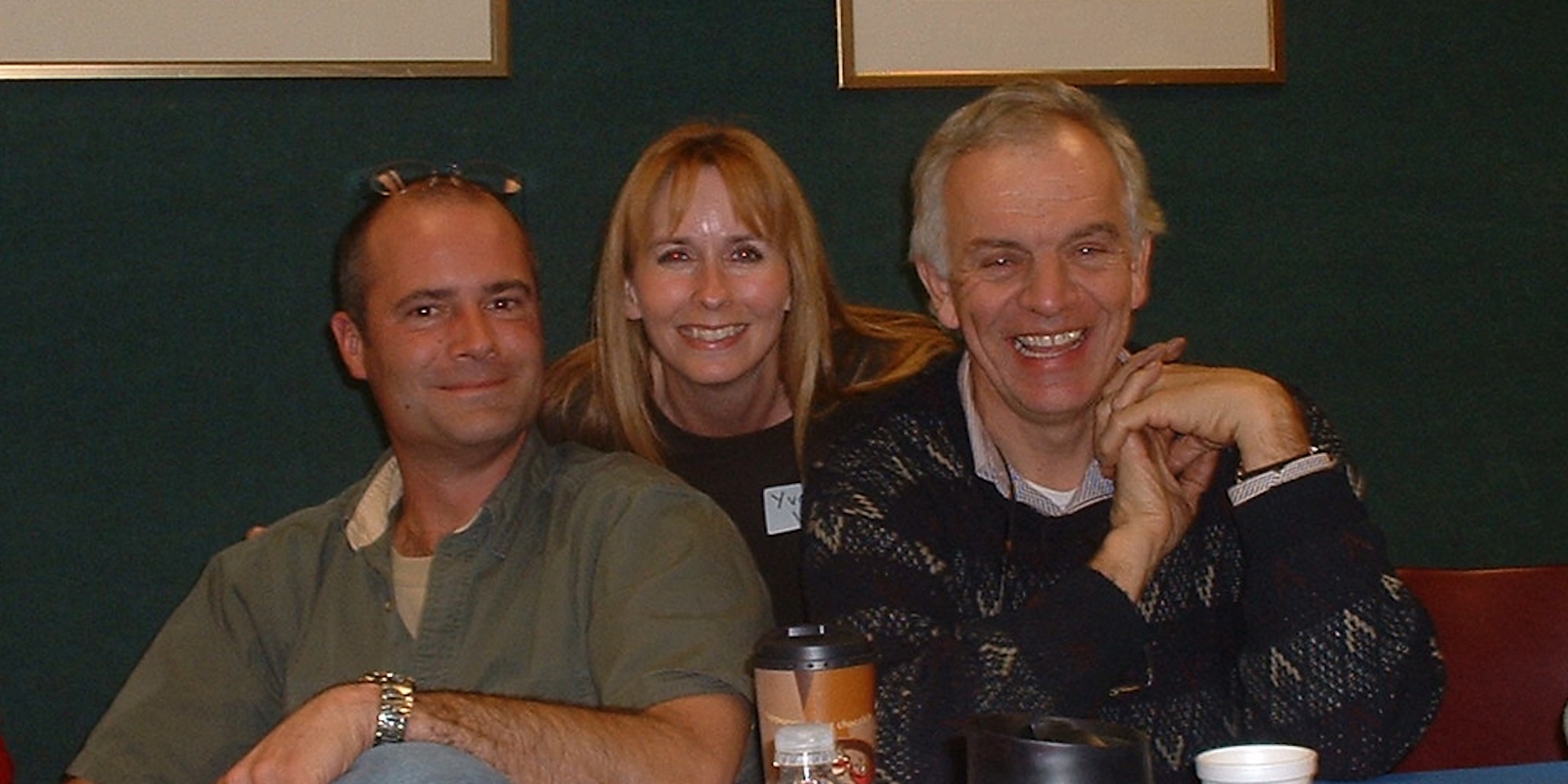
Dr. Robert Bowker, Cheryl Henderson, and Dr. Tomas Teskey in January 2007, conversing about the great mysteries of the hoof.
Bowker realized that previous work on hooves did not differentiate between healthy and unhealthy feet, so he set out to change that. In his cadaver studies, in horses under the age of 5, the back part of the foot looks the same. But after the age of 5, horses soon begin to split into two different groups that are easily identifiable. The good-footed horses developed fibro-cartilage in the digital cushion area of their hooves; while the bad-foot horses do not develop fibro-cartilage, and their digital cushions remain fatty connective tissue.
In the good-footed horses, the digital cushion is completely transformed into a very strong fibro-cartilage. It is clear that the horses were not born with this fibro-cartilage; rather, the tissue is stimulated (through movement), and the fibro-cartilage is created through this stimulation. Bowker is convinced that the horses’ early years are crucial for the development of the hoof, and only movement can create this crucial structure. Once the fibro-cartilage is created, it appears to be permanent. In addition to this fibro-cartilage in the digital cushion, a good foot also has thick lateral cartilages (3-4 times thicker), well-developed microvessels, and fibro-cartilage in the center of the frog.
A good foot will also tend to land slightly heel-first, which activates the proprioreceptors in the heel, stimulates the blood flow and allows for hemodynamic shock absorption. A toe-first landing is the sign of a sore horse.
A bad foot is not genetic; the horse was simply unable to adapt to improper load, negative stimulation, or inadequate environment. And, there is still hope for the older bad-footed horse. The cells to create the fibro-cartilage are still there, so the theory is that they can be activated through stimulation at any age, though no studies have yet confirmed this.
The Concept of Peripheral loading
Peripheral loading occurs when the edge of the hoof (hoof wall) bears more of the weight load. Peripheral loading always occurs with shoes, since they focus the weight upon the hoof wall. A shoe can therefore be called a peripheral loading device. However, peripheral loading can also occur with barefoot trimming, if the trim places more of the weight upon the wall. Over-trimming hoof structures such as the frog, sole and bars, so they have no possibility of sharing in the weightbearing load, will tend to create more peripheral loading. Allowing the hoof wall to grow too long will create more peripheral loading. Anytime the frog is not in contact with the ground, peripheral loading takes place. To make things more complicated, peripheral loading is completely dependent upon the hoof’s surface—a hard surface increases peripheral loading, while a softer surface decreases it. A solar plug (material packed into the hoof’s concavity) minimizes peripheral loading.
Peripheral loading is a negative situation for the hoof, because it severely interferes with blood flow inside the hoof. Bowker conducted experiments in velocity of blood flow in the hoof using Dopplar Ultrasound. What he discovered is that harder surfaces made the blood flow faster, and when it did that, it never “perfused” the tissues. It was just like a rainstorm in the desert (and I’ve seen plenty of those!)—water gushes down in a flood, but never sinks into the ground. The faster the blood flow, the less blood made it to the tissues!
“On softer surfaces (pea rock, sand or foam pads) blood flow will slow down and trickle through small vessels—microvenous vessels. On hard surfaces (cement or wood blocks), tissue perfusion dramatically decreases, so blood moves faster through foot—it must stay in the large vessels. Different surfaces will change tissue perfusion, with softer, more forgiving surfaces having the greatest tissue perfusion.”

One of Dr. Bowker’s sample coffin bones.
He also documented the effects of more extreme peripheral loading. With a shod hoof, blood flow actually came to a halt for a split second with every heartbeat, at the level of the horse’s fetlock. In a computer model that he developed with some engineers, he measured stresses on the coffin bone, and discovered that a peripheral load encouraged bone to be removed. A solar load encouraged bone to be laid down, or at least not removed.
The Responsiveness of the Hoof
The horse’s foot changes throughout its life. All four feet of a horse are different from each other, due to environment, exercise, trimming and active stimulation of the foot. And even wild horses’ hooves are all different, which makes it impossible to use them as a gold standard. There is no one wild horse model.
The horse’s foot is incredibly adaptive! It is only when its ability to adapt is exceeded, that lameness shows up. Amazingly enough, laminae are created in response to stress. The hoof is constantly adapting to stresses in the environment, and it appears that more laminae actually develop in the areas of stress inside the hoof. Fewer laminae are better, and extra laminae are signs of stress (they become thinner and longer, with a greater chance of laminitis). It has not yet been confirmed, but shod horses may have more laminae than barefoot horses. However, shod horses with toe clips do have more laminae around these clips. (This means we can mechanically increase the density of laminae!) Horses, in general, have more laminae on the flared side of their hooves, and more laminae at their toes. There are also more laminae in front of a sole callous; the pillar edge of a sole callous appears to be a stress area.
Not only are laminae created in response to stress, but it appears that hoof horn is, also. More about this later, but it appears that hoof tubules actually change direction according to load and stresses in the hoof wall.
The hoof is so responsive that there is a change in the physical contact area of the hoof between standing on concrete and standing on rubber. The hoof wall is fluid; hard but will actually move. There is a little more surface area of the hoof when the horse stands on hard rubber versus concrete, and this drastically reduces the pressure inside the hoof capsule. There is only 1/3 the amount of pressure on the hoof wall when standing on rubber versus standing on concrete.
The Necessity of Movement
Over and over again, we kept coming back to the importance of movement for the health of the foot. While this has long been a constant theme for natural hoof care, it was nice to finally have some science that backed this up! Bowker gave one clear reason why movement is so important: it improves the perfusion of the foot. This was readily measurable in his blood flow studies. Movement is also the only way the horse can develop the fibro-cartilage in the back of the foot, so a young horse without freedom of movement is a young horse destined to develop into a bad-footed horse!
Bowker has also taken his study of movement out into the field. Using very expensive and sophisticated pedometer devices, he measured the movement patterns of groups of horses living on 2-3 acre plots, and discovered that most healthy horses averaged about 4,000-6,000 steps per 24-hour period (3-5 miles). In contrast, horses living 24/7 in a stall took about 800 steps per day.
How the Hoof Grows
Walls: This seems obvious once you think about it, but for some reason, it had never been explored before Bowker’s studies. The hoof is shaped like a cone; the hoof is greater in mass distally than proximally. Either there are more cells at the bottom of the hoof, or the cells got bigger! Well, they didn’t get bigger. So Bowker wanted to find out how and why there were more cells at the bottom. He consulted with a specialist to record cell mitosis (cell division) in the hoof, and discovered there is no significant mitotic activity below the coronet. This is consistent with Dr. Chris Pollitt’s work. There are more cells, but where did those cells come from?
It turns out that the hoof wall does not simply grow straight down from the coronet. Most of it does originate from the coronet, especially the hard outer wall, but at least 1/3 of the wall is created from laminae-derived tubules. The secondary epidermal laminae carry little “grocery bags” of cells with them, and as they descend the hoof, these cells can go wherever the stresses are.
Bars: It has been previously assumed that the bars were structurally identical to the hoof wall—that they were simply an extension of the wall, turned around the frog. That doesn’t seem to be the case! The laminae of the bars are different than the laminae anywhere else. The bar’s laminae seem to be able to form tubular horn and contribute to both the wall and the sole. Bowker has been studying this on a microscopic level, and observed that tubules formed from the bars, grew forward, and migrated towards the toe. Bowker now believes that a significant amount of the sole might be coming directly from the bar laminae. Note: the bar laminae are contributing keratin cells to the sole, and so are the laminae (when well-attached). The solar corium does contribute sole, as well.
The Function of the Laminae
If the laminae are responsible for providing tubules to create at least 1/3 of the hoof wall, and the wall is fluid and dynamic, it starts to become questionable as to whether the laminae are actually a support structure for the hoof. Bowker now believes that the idea that laminae form the suspension that holds the horse up is false. He says that there is no direct connection that can be shown between the laminae and the coffin bone to indicate support. He compared it to saying that your hand is attached to your shoulder: true, but there is a lot of tissue in between!
Instead, Bowker theorizes that the function of the laminae is keratinocyte (keratin producing cells) storage, and producing tubules for the white line and sole.
Other Ideas
Pea rock: Horses (especially laminitic horses) love pea rock, which is a small, smooth, round river rock. Bowker recommends 3-6 inches of pea rock, on top of sand base. While horses may stand with their toes down in pea rock, their weight is actually on their heels. In his blood flow studies, pea rock created the highest blood perfusability rating.
Substance P Receptors: The sensory nerves in a horse’s foot are there for more than just pain; the nerves secrete a potent vasodilator called “Substance P.” Substance P acts on the small blood vessels in the foot. In the navicular horses he studied, the Substance P receptors are gone! The Substance P receptors were destroyed, and therefore there are less blood vessels and a loss of blood flow regulation. The decreased blood flow leads to remodeling of the bone. Navicular horses have lost the ability to control blood flow through the foot, due to the loss of these receptors. When a horse has lost the Substance P receptors, we end up in a catch-22 situation: the hoof can’t heal until blood flow improves, but the blood flow needs the Substance P vasodilator to improve!
Pulsating Veins: The veins in a horse pulsate. This is a unique feature of the horse, and something that has not been acknowledged before. “Veins of the distal limb of the horse have extensive musculature around them, and this smooth muscle appears under neural control. The vein pulsates like an artery!” However, in a laminitic horse, the venous pulses become less distinct and less consistent.
In regards to Dr. Bowker’s description, “The hoof wall inside is like peanut butter.” This one item probably got more comments of disbelief than any other, because so far no one has seen any peanut butter when dissecting a hoof. Not only was Bowker describing the inner wall, with its softer texture, but also I think he used this analogy to make a strong point: the hoof is changing, fluid and malleable, and NOT a rigid structure.
Everything we learned at Bowker’s presentation totally reverberated with us. We discovered new ways to think about the old familiar concepts and we acquired new tools to communicate these concepts to clients. How great is it to have an easily explainable scientific explanation for why shoes are best avoided? (just define peripheral loading!)
Trimming Ideas:
Note that Bowker is not a professional trimmer! His research does, however, provide us with a wealth of guidance. Trimming needs to be done as frequently as possible to minimize peripheral loading effects. Excess hoof growth will exacerbate peripheral loading. One of the best trimming tools for minimizing peripheral loading is the mustang roll, because it relieves the pressure on the outer hoof wall.
Bowker is a big advocate of backing up breakover on hooves: “Short toes are the best thing you can do to a foot!” His simple recommendations, which he calls the Physiological Trim, are short toes, short heels, and trimming for the 1/3-2/3 balance of the foot: 1/3 of the foot in front of the apex of the frog, and 2/3 behind it.
To create a thicker hoof wall, shorten the toes. To fix an underrun heel, shorten the toes. In an insulin-resistant horse, the inside of the hoof wall is actually unstable (keratinocytes are migrating more), so to compensate, reduce load on the hoof wall (short toes and mustang roll). Note: Bowker is against thinning the sole at the toe from the bottom, so flared toes should be backed up from the front only.
Clarification on bar trimming: Some people have taken Bowker’s recommendation of weight-bearing bars a little too much to heart, going so far as to discontinue all trimming of bars on all horses. I clarified this with him—when he talks about weight-bearing bars, he means the rear of the bar being weight-bearing upon impact. He said that he very much believes in concavity of the hoof! So if a bar needs trimming, it needs trimming. Bowker is, however, very much against excessive removal of the bars, i.e. trimming them to the point where they no longer function as a weight-bearing structure. The bars need contact with the ground for correct blood flow and sensory stimulation. (And remember, terrain matters, so a horse on deeper terrain would receive more contact with shorter bars, and vice versa.) Since the bars are responsible for some of the sole growth, they should not be removed. He also recommends that the soles should not become flat, but should have concavity, with a thicker sole if needed.
Believe it or not, there was much more, but we’ve run out of space in this article. In Bowker’s presentation, there is a great deal more detailed technical and scientific information, and photos, charts and data from many of his studies. He goes into much greater detail about his hoof growth and navicular studies. But don’t just take it from me, attend a Bowker presentation and evaluate his information first-hand.
by Yvonne Welz, published in The Horse’s Hoof Magazine, Issue 27, Spring/Summer 2007
See the full content listing of all issues of The Horse’s Hoof Magazine! We also provide instructions on how to read the issues for free on Hoof Help Online.
For a detailed listing of all articles on The Horse’s Hoof website, please visit our Article Directory.