An unrecognized reason for needlessly burying our horses??!!
Learn all about the signs of osteoporosis in horses’ coffin bones, direct from Dr. Bowker:
The demands placed upon the equine foot are greatest during loading of the limb and during certain disease states, such as laminitis, and over the years there has been extensive research effort towards understanding the biology of the foot during these movements and conditions.
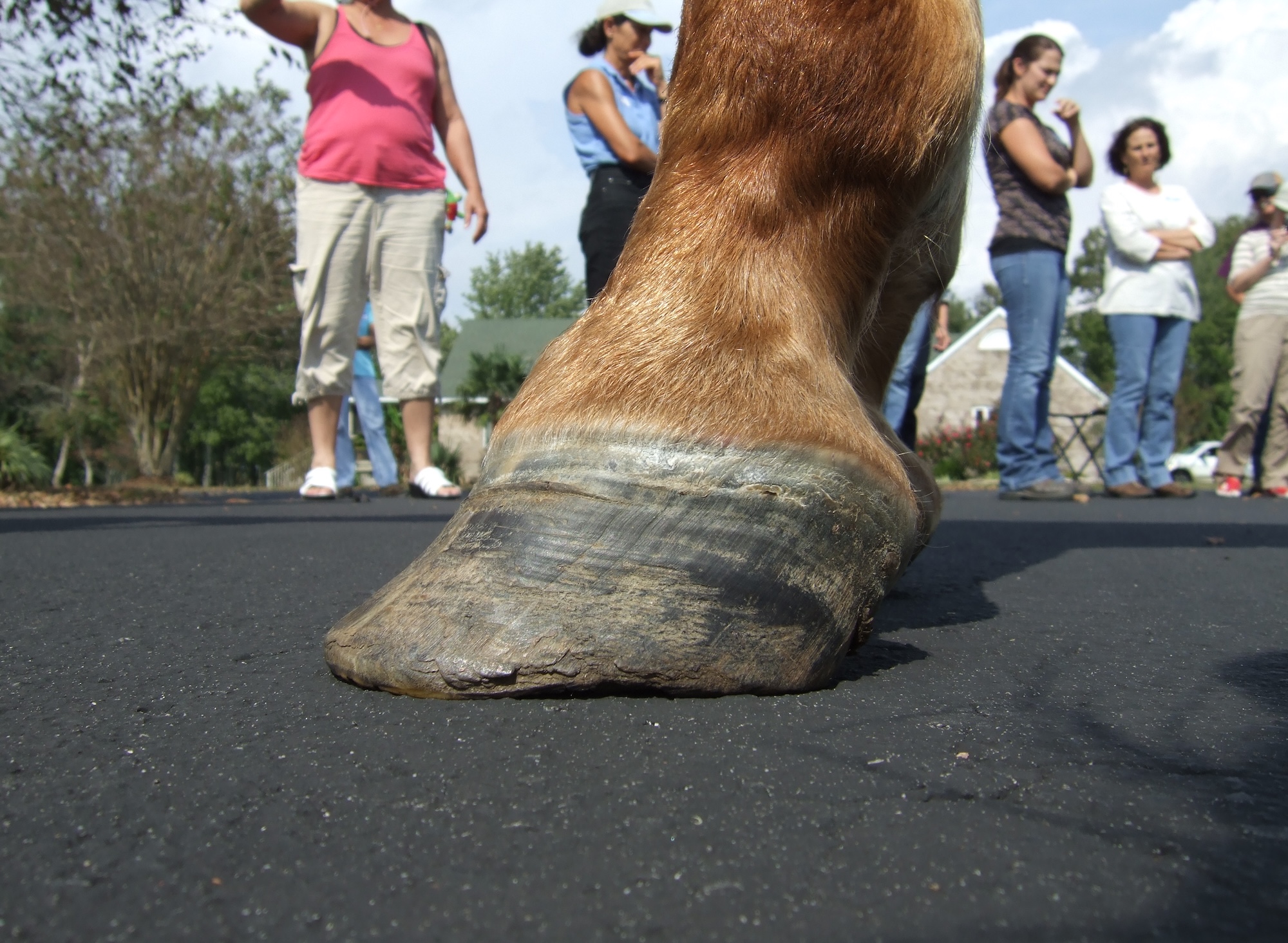
From “Equine Foot Biology 101,” our general understanding is that the hoof wall is the main support structure of the horse, and also provides protection for the more internal tissues, i.e. the coffin bone and palmar foot. Such a support mechanism occurs via the extensive inner lining of the hoof wall: the vertically-oriented sheets called epidermal laminae [lamina (singular) and laminae (plural) is Latin for “sheets”]. These laminae, consisting of approximately 600 vertical sheets, extend around the wall into the heels and bars along the solar surface and project towards the coffin bone. This anatomical arrangement of the inner hoof wall has long been known from the microscopic examination of the inner hoof wall in the previous, and most likely, earlier centuries.
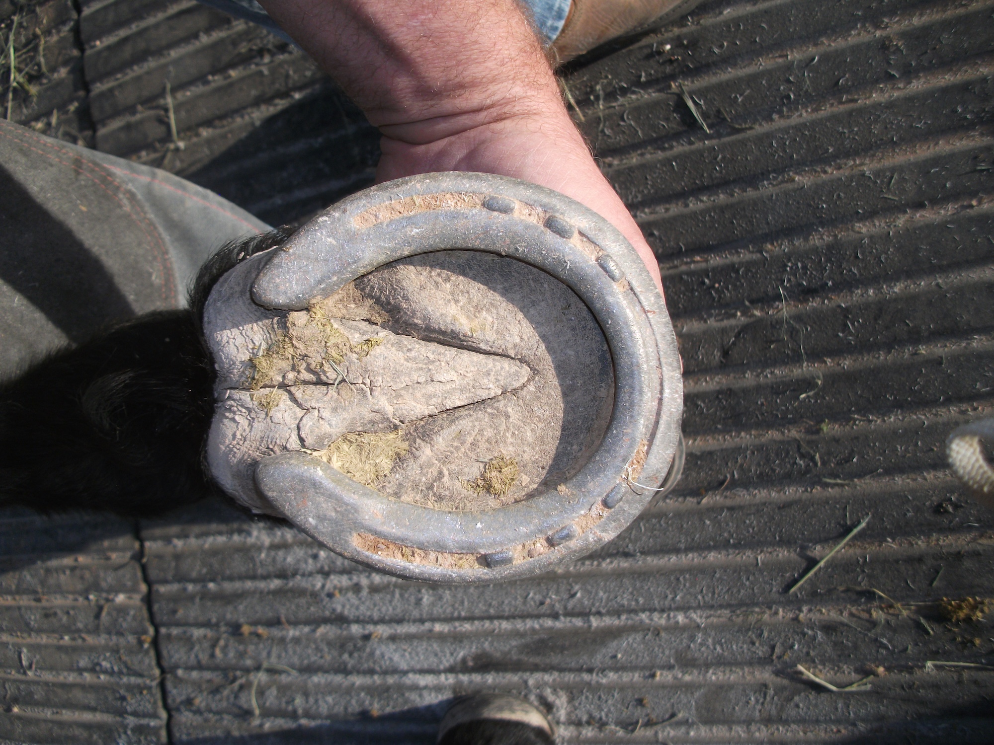
Such an anatomical arrangement provides a large surface area, seemingly to provide a primary function of support of the horse. This interpretation of the laminar function seems to have merit when the foot is peripherally-loaded, either with a shoe, or when the hoof wall extends a significant distance beyond the sole surface and the horse is standing or moving on a firm surface. Under these conditions, common sense tells us (and from imprints of a standing horse) that much or most all of the horse’s weight must be supported through the hoof wall when it contacts the ground surface (for example, 75-100%).
However, in the real world of many pastured domestic and of most feral horses, the hoof wall does not appear to significantly support the weight of the horse, but only a relatively small amount (i.e., 5-15% to 20%) due in part to the dirt plug present under their foot, or to the conformable surface (small rocks, lava rock, etc.) that they may be walking or standing on. Under these conditions, the majority of the weight is distributed over the sole via the earthen plug, with a much smaller percentage (for example, 5-15%) being placed upon the hoof wall. In these two extreme examples, the tissues of the foot will adapt to these loading paradigms, and their responses of each tissue will be different, as certain tissues will adapt and become THE primary support tissues, while other tissues will have a more secondary or perhaps even more tertiary support function.
Furthermore, the direction of the physical forces being applied to these tissues towards the foot tissues will be different in each instance: (1) with the peripherally-loaded foot, the forces will be more tangential to the inner foot tissues of the corium and particularly the coffin bone, while (2) in the second scenario (dirt plug/conformable surface), the physical forces of a solarly loaded foot will be more perpendicular to the corium and coffin bone, while a smaller tangential force (forces being applied to along the bone surface) will be applied to the dorsal cortical bone surface, serving as a secondary support tissue. How, and to what extent, these tissues respond will differ, and will depend upon the direction (and quantity) of the loading forces (vectors) being applied [i.e., forces being perpendicular to tissue (compression) or at an angle to the same tissue (strain and torsion) of the loads].
In terms of biomechanical stress, which is defined as load (weight) per unit area (load/ area), the tissues of the internal foot “sees” or experiences less of a load when the same load is spread over a larger area (i.e. sneaker mode), in contrast to a peripherally-loaded foot with a smaller surface area (i.e. high heel mode). In order to conceptualize the importance of load and surface contact area during weight-bearing in determining the biomechanical stresses within the foot, an illustration may help: for example, when you have a 1000 pound horse with approximately 60% of the load being on the forelimbs, it means that about 300 pounds are distributed to each forelimb (i.e. half of the 600 pounds). If the bottom of the horse’s foot is approximately 1 square foot for easy math calculations, such a contact area would result in the biomechanical stresses on the foot and the internal tissues being about 300 pounds per one sq ft.
On the other hand, if, in the same horse, the area of ground contact with the foot became smaller, such as, for example, ½ sq ft, when a shoe is applied to the foot, or the owner cleaned out the foot of a barefooted horse and walked the horse on a cement walkway, the effective biomechanical stresses on the foot and tissues would begin to increase, as now this load is 300 lbs per 1/2 sq ft or equivalent to about 600 lbs per one sq ft. Thus one can see that by just reducing the contact area, the stresses in the foot can increase.
Also, the direction of how the vector forces are applied to the foot (i.e. perpendicular vs. tangential) differs in these two examples, which, in turn, will effect how the hoof wall and bone tissues respond. Thus, one can see that this relative percentage of solar versus hoof wall can vary from foot fall to foot fall, depending upon whether the dirt plug falls out, or remains intact within the solar surface of the foot, and what their next step is, and what surface will they be stepping on, etc.! This relationship between area of support and the internal stresses within the foot is important, considering that horses spend much or most of their time standing around or walking, rather than running up and down hills and valleys!
These stresses are similar to wearing “sneakers” and “high heels,” and we are confident that most people will appreciate that the “sneaker-condition” is more comfortable than wearing high heels. With greater stress, internally the tissues will begin to be affected over time. In THH Issue 34, we wrote about the horse being “comfortable” (“Fingers, Frogs and Toes: Common Features”). Well, these biomechanical forces during loading are directly related to the stresses on internal tissues and to the “degrees of comfort”; generally with greater loading areas, the foot tissues have decreased stresses, but have greater comfort!
Now several questions come to mind regarding the above discussion, and the effects upon the coffin bone: 1) How is the coffin bone affected by these differences in biomechanical loading of the foot? 2) Do we have any evidence for such an idea of loading differences and effects upon the coffin bones? 3) Why do we think that it has importance in the horse?
We believe that by preferentially loading the solar surface, rather than loading the foot more peripherally via the hoof, more and more bone will be deposited within the coffin bone (or, more conservatively, will not be lost). This will result in greater bone density, which is better able to support the weight of the horse, and hence better to insure the long term health of the foot—rather than the opposite, of a gradual bone loss and eventually having to euthanize the horse, due to chronic foot problems. While this is just basic bone physiology—the so-called Wolfe’s law in action, we have some very preliminary evidence supporting this idea, (i.e. we choose to call the data preliminary, as we have only examined more than 300 coffin bones obtained from necropsy, and another group of horses involving all four coffin bones from more than 50 horses of known ages and breeds, and are slowly putting the data together.)

A brief summary is as follows: we have found that: 1) different bone densities exist in the coffin bones of horses; 2) a range of bone weights and densities of the coffin bones exist, with the forefeet having a wide range, as compared to a more narrow range of hind foot bones (Question: maybe this difference is related to more problems with the front feet?); and 3) the structural variations in coffin bone morphologies (weights and densities) may be related to the overall health of the foot (hoof, coffin bone and conformation) and of the horse. We are pretty confident of this idea, as age and breed and weight of horse do not appear to be factors in varying of bone densities (see example below)!
We believe that with time and proper foot support, more bone may be deposited within the coffin bone. Finally, a dense and structurally robust coffin bone is important in the overall health of the horse, as we do believe that significant bone loss does exist, and is not recognized in many of the foot problems in clinical practice! (NOTE: we realize that this is a bold statement, but we are coming to the conclusion that most horses examined at necropsy have varying degrees of osteoporosis in the coffin bone. In those horses with obvious foot problems, such as navicular syndrome, the coffin bones are severely osteoporotic (AAEP Proceedings, 2003). Furthermore, few “goodfooted” horses are presented at necropsy.)
Osteoporosis, a multifactorial disease in humans, is characterized by a reduction in bone mass and in the structural architecture of the bone. In the u.s., the disease is present in more than half of the people over 50 years of age, with a smaller percentage being affected clinically (i.e. microfractures and pain to obvious hip and limb fractures). (regarding horses, please reference “Bone Remodeling of the Equine Distal Limb” by Mark Fischer M.D. and Sheri Fischer R.N., B.S.N., in THH Issue 26.)
Bone undergoes constant remodeling of trabeculae and bone cortices throughout the life of the organism, due to both “proper” and “improper” loading, along with hormonal influences (Wolfe’s Law). With “proper” loading our bones remain straight, robust and healthy. In the horse, the coffin bone also continues to remodel, and we believe should be balanced to produce a symmetrical foot and internal coffin bone with good structural support. Although most trimmers and farriers would agree with this statement, one can appreciate that the asymmetric foot (steep and flared sides) is far too common when examining feet, suggesting that a balanced foot may be an uncommon occurrence?
When the loading becomes asymmetrical (or uneven balance), then both the hoof wall and the bone begin to adapt and respond, by depositing more horn and bone on one side of the foot, and actually removing horn and bone from other side of foot. such an imbalanced loading and unloading of the bone will eventually become “clinically significant,” as the cortical and trabecular bone become less dense on one side of the foot, until it can no longer support the load of the horse, especially during exercise or workouts.
Historically in women, it was believed that when the bone was weakened by significant bone loss, injury occurred when the woman fell, causing the fractured hip joint. However, more recent evidence suggests that microfractures or microscopic fractures through the cortices and the trabeculae are present first and actually contribute to the injury, as the bone is no longer able to support the person’s weight, and hence the woman falls, creating even more fractures that can now be seen using radiographs. We hypothesize that a similar process (i.e. microfractures) is occurring in the forefeet and coffin bones of domestic horses, and is at present unrecognized, unless one is aware of the more intricate details of the coffin bone structure. We are presently trying to develop a field procedure for detecting this condition in live horses.


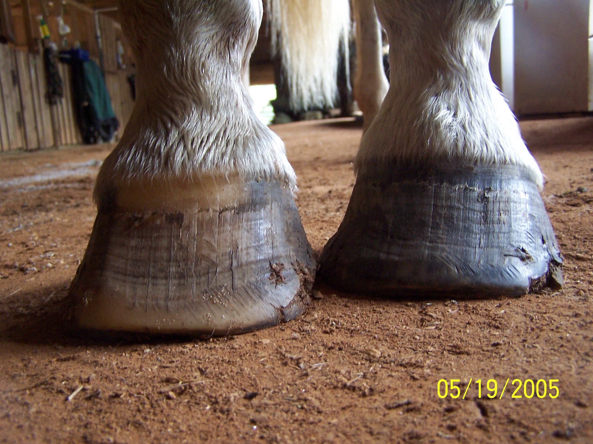
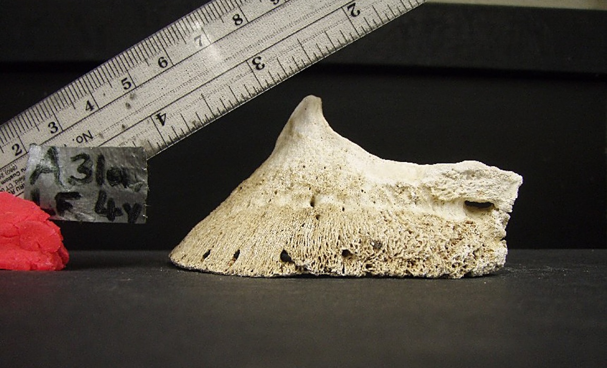
In the two lateral views of these apparently “normal” coffin bones (photos above), they are considered to be reasonably good coffin bones, as they have greater density and fewer changes than the majority of bones. For the purposes of illustration, we are showing that this 4 yr old coffin bone is less dense than the 31 yr old coffin bone, and would likely have clinical problems later in life. We have found at least five different “descriptive markers” on the bones that we use as “indicators” of how much bone loss there may be in these feet: the more markers that are present, the greater degree of bone loss we are finding. We are initially examining feet of horses of known ages, breeds, etc., by radiographing them, describing and measuring external hoof features and then harvesting the bones and examining them histologically, and trying to correlate the hoof observations with the radiographic information and the coffin bone morphological descriptors, as well as with the history of the horse, if possible. Obviously, a very long and tedious process, but we are proceeding forward with this goal.
Today, we will briefly present one of these markers—the changes in the palmar processes (PP)—and show you why we believe that osteoporosis is a common but unrecognized occurrence in the general horse population, and that the structural support of the coffin bone is critically important to a healthy foot. As we have mentioned before, the elongation of the palmar processes is analogous to putting “outriggers” on a small boat, as one attempts to stabilize any uneven loading that may exist. In the horse’s foot, we think that the coffin bone is also attempting “to balance and stabilize itself.” All changes in the PP that we have found to date indicate greater problems with the foot and greater degrees of osteoporosis of the coffin bone: increased porosity (greater number and size) of the PP (normally very small micropores are present); increased length beyond 1.5-2.0 cm upwards to more that 3.0 cm from the coffin joint collateral ligament (normally want a short <1.0-1.5 cm length of PP); contributions to the ossified lateral cartilage; microscopic cortical bone thinning; and increased trabecular spacing, to name a few. These changes merely are indicative of progressive bone loss in the coffin bone in horses.
In these two photographs above, one (4 yr old LF) has a greater porosity in the bone (more micropores, with some being up to a mm wide) and has less bone density than coffin bone of the 31 yr old horse. When similar bones having these changes in the PP are examined under the microscope, the PP has more osteoporotic bone with a thin cortex and increased trabecular spacing of the PP bone, as well as in other areas of the coffin bone (see photo below). Clinical relevancy of active and potentially pathological remodeling may be occurring when positive hoof tester responses are present over heels. These increased porosity bone changes are evident in routine radiographs, indicating that such remodeling changes are extremely common in the domestic horse in the United States. Future study should help shed some light on these questions regarding the coffin bones of horses!

These changes we are beginning to see in the coffin bones merely show that we have much to learn about the horse’s foot, even though we have made great strides during the past few decades. That is why I often use the lines of Robert Frost, the New England poet, from the poem “Stopping By Woods On a Snowy Evening” to indicate some of our goals:
“And miles to go before I sleep, And miles to go before I sleep.“
There’s lots to do!!!
About the authors: Dr. Robert Bowker has spent the past few years studying the functioning of the equine foot in health and disease at Michigan State University, and is now working on his rehab center – Corona Vista Equine Center in Michigan. Ms. Tara Calvert-Jackson is pursuing studies this fall at MSU, and will actively be studying and researching the foot.
by Dr. Robert M Bowker and Tara Calvert-Jackson, published in The Horse’s Hoof Magazine, Issue 35, Summer 2009
See the full content listing of all issues of The Horse’s Hoof Magazine! We also provide instructions on how to read the issues for free on Hoof Help Online.
For a detailed listing of all articles on The Horse’s Hoof website, please visit our Article Directory.